
1. Introduction
Cell culture remains a foundational technology across biomedical research, biotechnology, disease modelling and drug discovery. Conventional two-dimensional (2D) monolayers continue to offer important advantages. They are comparatively simple to establish, scalable, accessible, and compatible with a wide range of molecular and cellular assays. In contrast to the structure, extracellular-matrix interactions, and gradients found in tissues, cells grown on rigid planar surfaces encounter an artificial spatial environment. [1-3]
The development of three-dimensional (3D) models, including as spheroids, organoids, scaffold-based systems, microfluidic platforms, and engineered tissues, has accelerated due to this constraint. These methods increase researchers' experimental toolkit rather than taking the place of 2D culture. The most appropriate model depends on the biological question, cell type, experimental endpoint and required level of physiological complexity. [1-4]
2. 2D Culture: Practical, scalable and still relevant
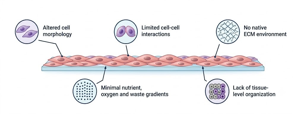
A 2D monolayer continues to be an effective experimental system for many research applications. Routine cell expansion, transfection, viability assays, Western blotting, gene-expression studies, and many screening applications can be performed efficiently using conventional culture. Additionally, microscopy, sampling, and experimental standardization are made easier by the relative simplicity of 2D systems.
Nevertheless, planar growth can alter cell morphology and polarity and may not reproduce complex cell–cell or cell–matrix interactions. It also provides a less representative representation of spatial gradients than many 3D systems. These differences can become particularly important when the phenotype under investigation depends on tissue architecture or microenvironmental cues. [2,3]
3. Why 3D models are gaining importance
3D culture allows cells to organize within a spatial environment that can better reproduce selected features of tissue architecture. Depending on the platform, researchers can investigate cell–cell interactions, extracellular-matrix signalling, diffusion gradients and spatially heterogeneous responses to treatment. [1,3,4]
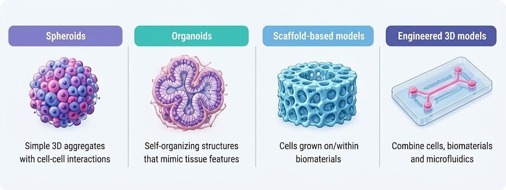
• Spheroids: useful for studying cellular aggregation, diffusion gradients and multicellular responses.
• Organoids: self-organizing models that reproduce selected structural and functional characteristics of
tissues.
• Scaffold-based systems: provide defined structural support for cellular organization.
• Microfluidic and engineered models: enable controlled environmental conditions and integration of multiple
biological or engineering components.
In the fields of disease modelling and drug discovery, where traditional monolayers could miss crucial facets of tissue behaviour, the growing complexity of 3D models is especially pertinent. Increased complexity also brings in new factors that need to be verified and regulated [3,5].
3.1. Key takeaway
The direction of modern cell culture is not a simple transition from 2D to 3D. It is a move toward selecting the experimental model that best matches the scientific question.
4. Organoids and advanced tissue models
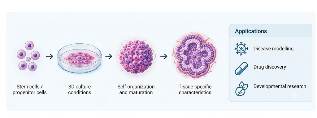
Organoids have emerged as important 3D experimental models because they can self-organize under defined conditions and reproduce selected features of their tissue of origin. Intestinal, cerebral, hepatic, renal, retinal, and other organoid systems are increasingly used to study development, disease mechanisms, tissue responses, and therapeutic interventions [4,6]. In oncology, organoids can retain selected characteristics of patient tumours, providing valuable models for investigating tumour heterogeneity, treatment response, and therapeutic resistance, while the integration of immune and stromal components is further enhancing their biological relevance [7,8].
4.1 From organoids to more complex systems
Next-generation in-vitro models increasingly integrate cellular self-organization with engineering approaches such as biomaterials, hydrogels, microfluidics, bioprinting, and organ-on-chip technologies. These platforms can better control spatial organization, extracellular-matrix interactions, nutrient delivery, and mechanical cues, providing more physiologically relevant environments for studying complex biological processes that are difficult to reproduce in conventional monolayer culture [3,9].
4.2. The reproducibility challenge
Greater model complexity does not necessarily ensure greater experimental reliability. Variability in cell source, matrix, medium, culture conditions, and analytical methods can affect 3D and organoid models, making standardization and quality control essential for reproducible research [6,10].
5. Cell culture medium: The experimental environment
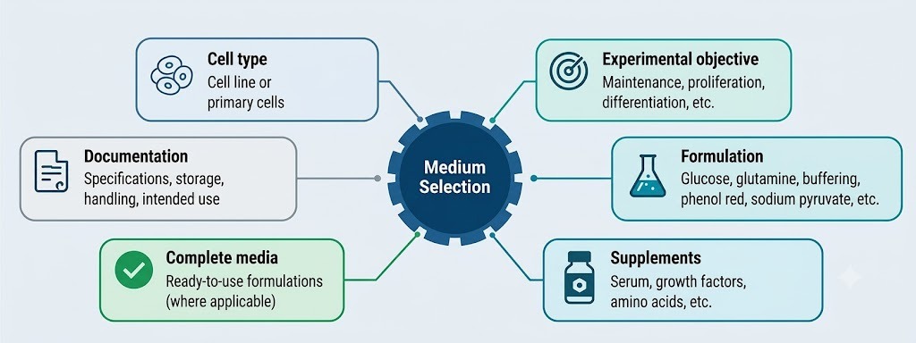
The culture medium is not simply a nutrient solution; it is a major component of the cellular microenvironment. Basal formulations generally provide amino acids, vitamins, salts, glucose and buffering components, while supplements can supply growth factors, hormones, lipids and other factors required by specific cell systems. The composition of the medium can influence cell growth, phenotype, metabolism and experimental readouts [11,12].
Common basal media include DMEM, RPMI-1640, MEM, MEMα and DMEM/F-12. However, the most familiar formulation is not necessarily the most appropriate one. Medium selection should be driven by the cell type, intended application, culture format and experimental objective.
5.1 A Practical Selection Framework
• Cell identity and source: Confirm the recommended conditions for the specific cell line, primary cells or
stem-cell-derived model.
• Experimental objective: Distinguish between maintenance, expansion, differentiation, functional assays and
specialized applications.
• Formulation: Evaluate glucose, glutamine, buffering components, sodium pyruvate, phenol red and other
relevant constituents.
• Supplements: Determine the need for serum, growth factors or defined additives.
• Model architecture: Recognize that 2D monolayers, spheroids and organoids may have different nutritional
requirements.
• Documentation: Record formulation, lot information, supplements, storage and culture parameters to
support reproducibility.
5.2 Serum, serum reduction and defined culture systems
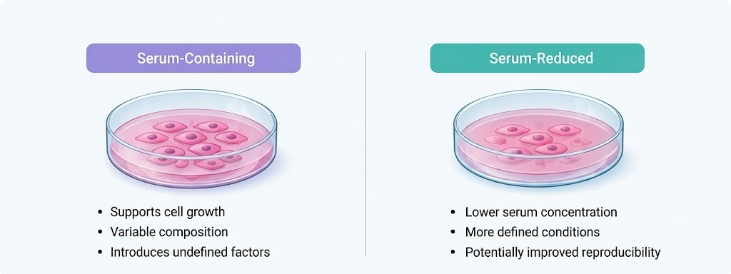
Fetal bovine serum (FBS) is widely used as a complex source of growth-promoting factors in cell culture. However, its biological complexity and substantial lot-to-lot variability can influence cell growth and experimental outcomes, introducing an important source of uncontrolled variation [12-15]. For applications requiring greater control of the extracellular environment, serum-reduced, serum-free, or chemically defined systems may offer suitable alternatives, although their selection should be guided by the specific cell type and experimental objective and validated under the intended culture conditions [13,14].
5.3 Why medium consistency matters
Variability in raw materials and media components can affect cell growth, viability and functional characteristics. This is especially relevant in translational research and bioprocessing, where reproducibility and process consistency are critical [12]. Consequently, researchers should treat medium selection as an experimental variable rather than a routine purchasing decision. A formulation should be evaluated according to its intended application, documented carefully and validated under the laboratory's specific conditions.
Looking for cell culture media for your research workflow?
Explore basal media, specialized formulations, reduced-serum solutions and complete cell culture media from Universal Biotechnology.
Explore Cell Culture Products →
6. Universal Biotechnology: Cell culture solutions for research
Universal Biotechnology provides a diverse portfolio of cell culture media, supplements, and specialized formulations designed to support a broad range of research applications. The portfolio encompasses:
• Conventional basal media
• Specialized formulations
• Reduced-serum culture solutions
• Complete media systems for selected cell types
This range enables researchers to select culture conditions based on the biological requirements of the model and the specific objectives of the experiment.
6.1 SR DMEM/F-12 (1:1) and SR Buffer
For research applications requiring reduced-serum conditions, Universal Biotechnology offers SR DMEM/F-12 (1:1) as a basal medium formulated to support reduced-serum culture workflows. According to the manufacturer's product information, the formulation has been evaluated for parameters including cell proliferation, cellular morphology, and genome stability under specified testing conditions. SR Buffer is also available for use with appropriate basal or complex media in reduced-serum culture systems.
As serum requirements and cellular responses can vary considerably between models, formulation performance should be assessed in the context of the specific cell type, culture conditions, and experimental endpoint. Appropriate laboratory validation remains essential before implementation in experimental workflows.
6.2 Procell System: Supporting diverse cell culture applications
The Procell System portfolio includes widely used basal formulations such as DMEM, RPMI-1640, and MEMα, together with specialized media including Neurobasal Medium and TNM-FH Medium. The portfolio also includes complete media formulations for selected cell systems, including Caco-2, BV2, Hep G2, SH-SY5Y, and NK-92, as well as MEMα without nucleoside for applications involving DG44 and other DHFR-deficient cell systems.
A specialized Nanobacteria Removal Medium is also included within the portfolio for specific culture-contamination management applications. Its use should be determined according to the requirements of the experimental system and the manufacturer's recommended conditions.
6.3 Matching medium to model complexity
As cell-culture models progress from conventional monolayers to spheroids, organoids, and engineered 3D systems, medium selection becomes increasingly critical. A formulation optimized for a 2D monolayer may not necessarily provide the nutritional and signalling environment required for a more complex 3D model. Medium composition can interact with extracellular-matrix conditions, growth-factor signalling, cellular metabolism, and cell–cell communication, thereby influencing phenotype and experimental outcomes.
Therefore, medium selection should be application-driven rather than based solely on formulation familiarity or product category. The objective is to establish a reproducible combination of cell source, medium, supplements, matrix, and culture conditions that maintains the desired cellular phenotype and generates consistent experimental readouts. Careful optimization, documentation, and validation of these parameters are particularly important for advanced and translational cell-culture workflows.
7. Designing a reproducible cell-culture workflow
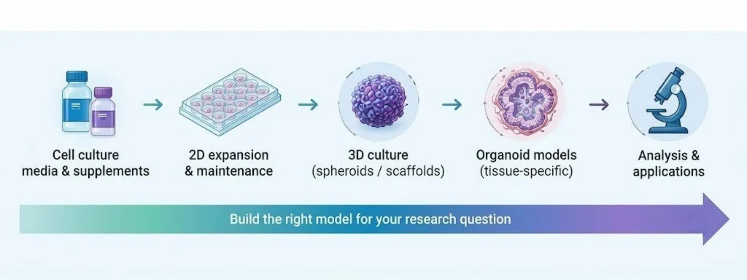
A robust cell-culture workflow begins with the scientific question and works backward to the experimental model. Choosing a more sophisticated platform is justified when the added biological complexity provides information that cannot be obtained reliably from a simpler system.
7.1 A Researcher’s decision framework
• Define the biological question and primary experimental endpoint.
• Select the most appropriate cell source and culture architecture.
• Establish the recommended basal medium and supplement requirements.
• Control critical variables such as passage number, seeding density, matrix conditions, incubation
parameters and medium changes.
• Document reagent identity, lot information and storage conditions.
• Define quality-control readouts appropriate to the model.
• Validate phenotype and function before using the system for comparative experiments or screening.
7. 2 D and 3D Are complementary platforms
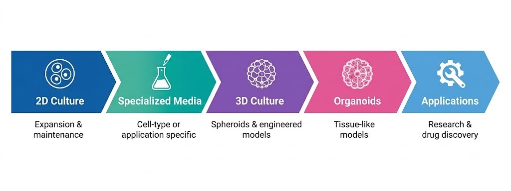
A practical workflow may use 2D culture for expansion and routine characterization before transferring cells into a 3D model for the experimental phase. This staged approach can reduce unnecessary complexity while allowing researchers to introduce tissue-relevant architecture when it is scientifically justified. For example, conventional monolayers may be appropriate for routine proliferation or molecular assays, whereas spheroids may be selected when cell aggregation and diffusion gradients are central to the question. Organoids may be more appropriate when tissue-specific organization or disease modelling is required. Engineered or microfluidic systems can be considered when controlled physical or biochemical microenvironments are essential [3,5,9].
8. Emerging trends in cell culture
8.1 Automation and high throughput 3D culture
The increasing use of 3D models has created a parallel need for automation, imaging and quantitative analysis. Traditional 3D workflows can be labour intensive and may be difficult to integrate into conventional high-throughput screening systems. Advances in microfabrication, automated culture, high-content imaging and computational analysis are helping address these limitations [5].
8.2 Engineering tissue-relevant microenvironments
Biomaterials, microfluidics and bioprinting are providing researchers with greater control over the physical and spatial characteristics of cell culture systems. These technologies can be used to organize multiple cell types, generate defined architectures and reproduce selected aspects of tissue mechanics and transport [9].
8.3 Toward more human relevant models
The broader direction of cell culture research is toward models that provide greater biological relevance while maintaining experimental control and reproducibility. Organoid systems, organ-on-chip platforms and engineered tissues are important components of this transition, but their value ultimately depends on appropriate validation against the biological question they are intended to address [6-10].
8.4 Research-Use Considerations
The products discussed in this article are presented for laboratory research applications. Product-specific intended-use statements, specifications, storage requirements and handling instructions should be reviewed before use. Research Use Only (RUO) products are intended for research applications and are not represented for use in clinical diagnostic procedures.
9. Conclusion
Cell culture in 2026 is best understood as a spectrum of experimental models rather than a choice between old and new technologies. Conventional 2D culture remains highly valuable for many applications, while 3D models provide additional opportunities to investigate tissue organization, cellular interactions and disease biology. The central challenge is to match model complexity with scientific need.
Across both conventional and advanced systems, the quality of the culture environment remains fundamental. Appropriate medium selection, controlled culture conditions, documentation and validation are essential for reproducible research. As cell culture moves toward increasingly sophisticated models, the ability to combine biological relevance with experimental consistency will remain a defining requirement of high-quality in-vitro research.
Have a specific cell-culture requirement?
Connect with the Universal Biotechnology team to discuss suitable media, supplements and research-use formulations.
Contact Universal Biotechnology →
References
1. Edmondson R, Broglie JJ, Adcock AF, Yang L. Three-dimensional cell culture systems and their applications
in drug discovery and cell-based biosensors. Assay Drug Dev Technol. 2014;12(4):207–218.
2. Duval K, Grover H, Han LH, et al. Modeling physiological events in 2D vs. 3D cell culture.
Physiology (Bethesda). 2017;32(4):266–277.
3. Badr-Eldin SM, Aldawsari HM, Kotta S, et al. Three-dimensional in vitro cell culture models for efficient
drug discovery: progress so far and future prospects. Pharmaceuticals. 2022;15(8):926.
4. Lee SY, Koo IS, Hwang HJ, Lee DW. In vitro three-dimensional (3D) cell culture tools for spheroid
and organoid models. SLAS Discov. 2023;28(4):119–137.
5. Fitzgerald KA, Malhotra M, Curtin CM, O’Brien FJ, O’Driscoll CM. Life in 3D is never flat: 3D cell culture for
drug discovery and development. Adv Drug Deliv Rev. 2022;175:113775.
6. Blatchley MR, Anseth KS. Middle-out methods for spatiotemporal tissue engineering of organoids. Nat Rev
Bioeng. 2023;1:329–345.
7. Polak R, Zhang ET, Kuo CJ. Cancer organoids 2.0: modelling the complexity of the tumour immune
microenvironment. Nat Rev Cancer. 2024;24:523–539.
8. Marx V. Closing in on cancer heterogeneity with organoids. Nat Methods. 2024;21:551–554.
9. Huang MS, Christakopoulos F, Roth JG, et al. Organoid bioprinting: from cells to functional tissues. Nat Rev
Bioeng. 2025;3:126–142.
10. Lee, H., Yang, S., Lee, K.J., Kim, S.N., Jeong, J.S., Kim, K.Y., Jung, C.R., Jeon, S., Kwon, D., Lee, S.
and Lee, H., 2024. Standardization and quality assessment for human intestinal organoids.
Frontiers in cell and developmental biology, 12, p.1383893.
11. Yao T, Asayama Y. Animal-cell culture media: History, characteristics, and current issues. Reprod Med
Biol. 2017;16:99–117.
12. Bones J, Mittermayr S, O'Callaghan P, et al. Examining the sources of variability in cell culture media used for
biopharmaceutical production. Biotechnol Lett. 2017;39:1401–1410.
13. van der Valk J, Bieback K, Buta C, et al. Fetal bovine serum (FBS): past – present – future.
ALTEX. 2018;35(1):99–118.
14. van der Valk J, Brunner D, De Smet K, et al. Optimization of chemically defined cell culture media—replacing
fetal bovine serum in mammalian in vitro methods. Toxicol In Vitro. 2010;24(4):1053–1063.
15. Brunner D, Frank J, Blättler T, et al. Serum-free cell culture: the serum-free media interactive online database.
ALTEX. 2010;27(1):53–62.